What does HistoIndex do?
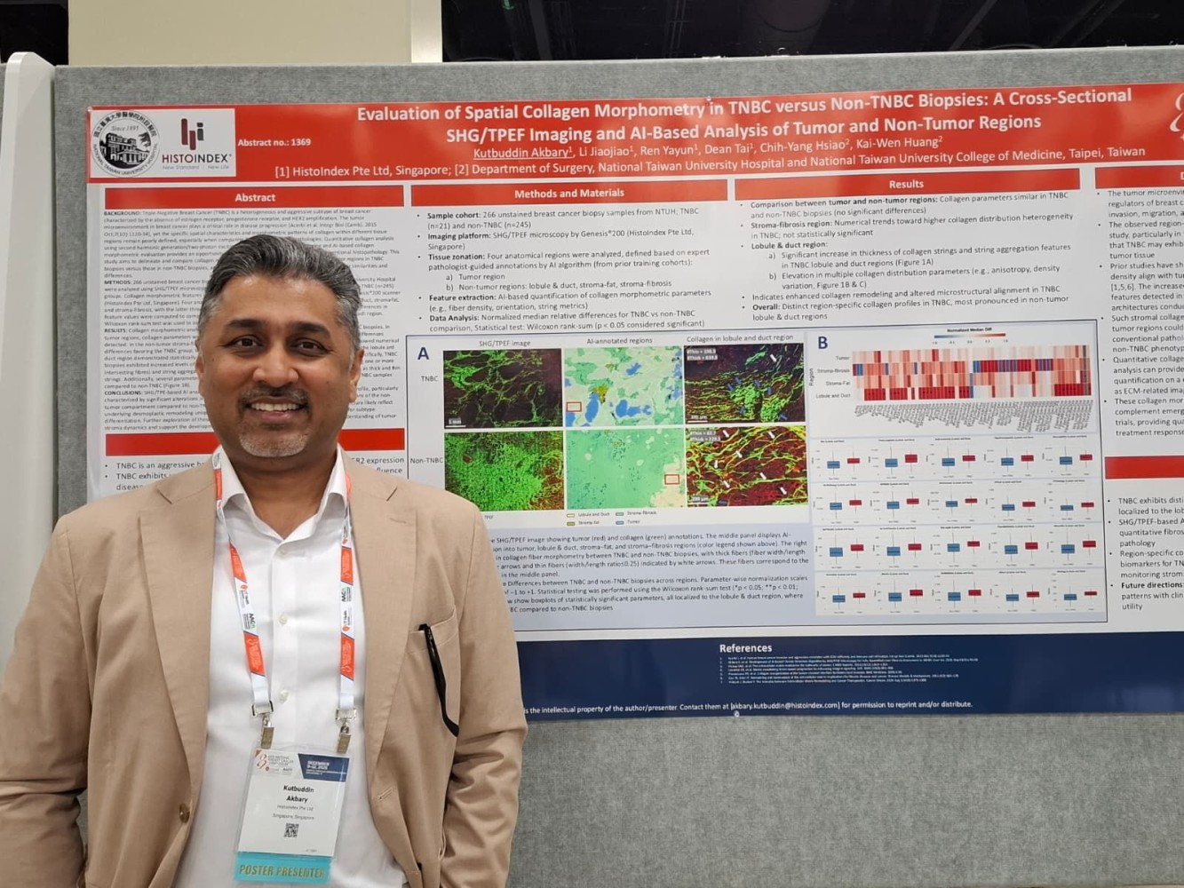
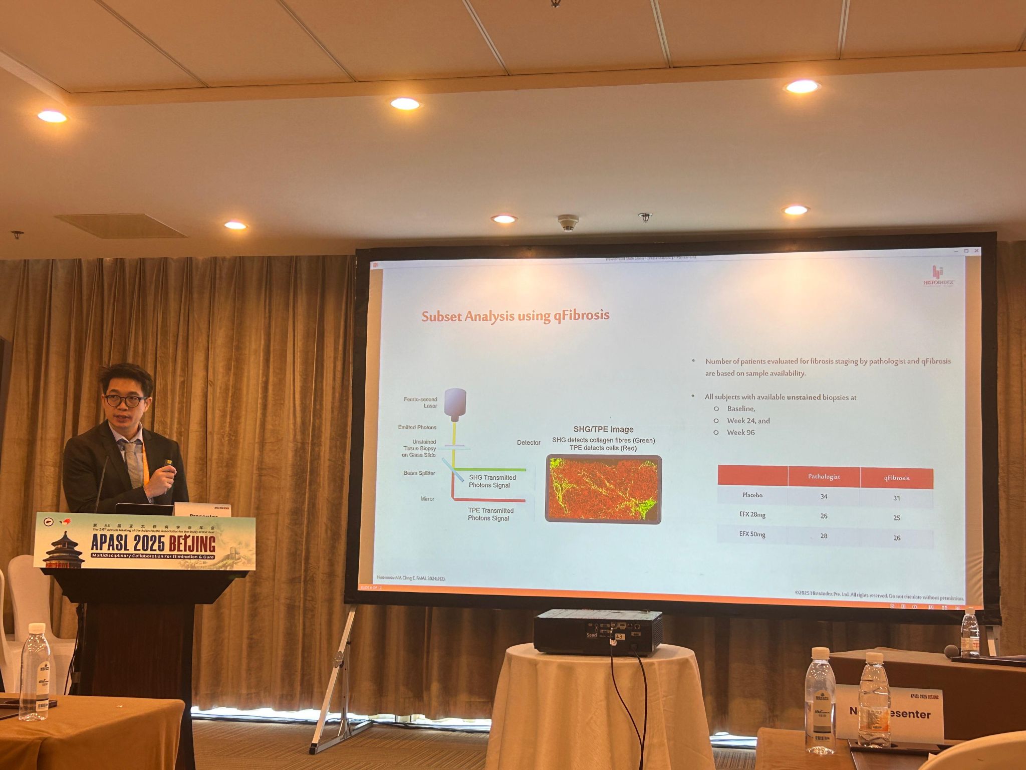
HistoIndex is a leader in AI-powered digital pathology and stain-free imaging technology that quantifies disease features, such as fibrosis in liver tissue, with objective, reproducible data. Our platform combines advanced imaging with explainable AI to support research, clinical trials, and clinical diagnostics.
What is digital pathology and how does HistoIndex use it?
Digital pathology involves scanning glass slides into high-resolution digital whole slide images (WSI) and applying computational analytics. HistoIndex uses AI-driven algorithms to analyze these images, enabling precise quantification of histological features and reducing subjective variability that occurs with manual microscopy.
What is stain-free imaging and why is it important?
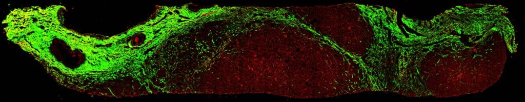
Stain-free imaging, such as Second Harmonic Generation (SHG) microscopy, visualizes tissue without chemical stains, capturing intrinsic signals from biological structures. This method produces consistent imaging data that can be quantitatively analyzed for morphological features like collagen in fibrosis, improving accuracy over traditional stains.
What diseases or conditions can HistoIndex help analyze?
While HistoIndex is widely known for liver fibrosis evaluation, including MASH (Metabolic dysfunction-associated steatohepatitis), its technologies also support broader research into fibrotic diseases and tissue architecture analysis relevant to drug development and diagnostic pipelines.
How does AI enhance traditional histology analysis?
Artificial intelligence augments histology by identifying subtle patterns and features in tissue that may be difficult for the human eye to detect, delivering quantitative metrics rather than subjective scores, increasing consistency and accuracy in analysis.
Can HistoIndex services support global research collaborations?
Yes, the digital pathology platform supports remote data transfer and analysis, facilitating collaborations across geographic regions and enabling researchers and clinicians worldwide to access robust quantitative histology data.
How does HistoIndex’s technology improve clinical decision-making?
By transforming complex histological images into objective, reproducible measurements, HistoIndex supports data-driven interpretations, enhancing diagnostic confidence and offering deeper insights into disease progression or therapeutic impact.
Is HistoIndex’s AI pathology platform validated by scientific research?
HistoIndex and its proprietary metrics such as qFibrosis® have been featured in peer-reviewed studies and scientific publications demonstrating quantitative digital pathology’s efficacy and reproducibility in disease assessment.