
Digital Pathology: The Future of Diagnosis
Driven by AI
Digital pathology refers to the digitalization of glass slides for computational analysis. It involves scanning tissue sections at high resolution to create whole slide images (WSI) that can be stored, managed, viewed, analyzed and shared digitally. Artificial intelligence provides cutting-edge analytics to maximize the findings from digital pathology images.

Transforming Assessments with Digital Pathology
Beyond the Microscope
Digital pathology allows for digitisation of tissues slides for visualisation, and allows for quantification of features when coupled with image analysis software and computational algorithms in an automated and objective manner. Importantly, this removes the subjectivity of manual staging/scoring and instead generates quantitative metrics.
How AI Uncovers Hidden Disease Clues
Quantifying the Invisible with AI
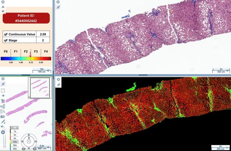
Benefits of AI in Digital Pathology
- Characterization and quantification of the morphology, architecture and spatial distribution of histopathological structures, cell types and features
- Provides continuous values instead of ordinal stages/grades
- Captures subtle patterns and changes indicative of treatment response or disease progression machine-learning algorithms
- Eliminates intra-observer and interobserver variability in assessments
- Facilitates efficiency gains in high volume clinical trials and routine assessment
AI Digital Pathology in Liver Disease Drug Development
Accelerating Advancements
Digital Pathology FAQs
Digital pathology is the practice of capturing high-resolution whole slide images (WSI) of traditional glass slides and using computer-based tools to view, manage, and analyze them digitally, enabling remote review, advanced analytics, and data-driven interpretation.
Unlike viewing tissue samples through a microscope, digital pathology digitizes entire slides, allowing images to be stored, shared, annotated, and analyzed with software and AI, reducing subjectivity and variability compared to manual interpretation.
Digital pathology paired with AI analytics uncovers subtle patterns in tissue biopsies, such as collagen architecture in fibrosis, providing more objective and reproducible data than traditional semiquantitative scoring. This enhances sensitivity for detecting disease progression or treatment response in clinical and preclinical studies.
Artificial intelligence (AI) algorithms analyze digital slide images to quantify morphology, architecture, and spatial distribution of histological features, identifying subtle disease signals that may be difficult for the human eye to detect consistently.
Digital pathology provides:
- Objective quantitative analysis of histological features.
- Improved collaboration and remote review by enabling digital sharing of slides.
- Enhanced workflow efficiency and data management, with easier storage and retrieval of digital slides.
- Reduced variability between observers.
Yes, by delivering consistent, data-driven insights from tissue images, digital pathology supports more reliable assessments that can influence patient management strategies, especially when combined with AI for complex pattern recognition.
Yes, it’s increasingly used in clinical and research settings to quantify treatment effects, generate reproducible endpoints, and support regulatory submissions in drug development, especially for diseases like fibrosis where precise measurement is critical.
Digitized slides can be shared electronically across institutions, enabling experts to review and consult on cases from different locations, enhancing access to specialist insights and second opinions.
Yes, digital pathology workflows typically require slide scanners to convert glass slides into whole slide images, as well as software platforms for viewing, analyzing, and managing these digital files.