Introduction
In the evolving landscape of medical research and diagnostics, histopathology plays a pivotal role in the accurate assessment of diseases like cancer and fibrosis. Histopathology, the study of tissue changes caused by disease, depends heavily on imaging techniques to visualize the intricate structures within tissues. These techniques, whether traditional or innovative, are essential in evaluating disease progression, especially in liver conditions such as fibrosis. Accurate imaging is critical in ensuring that clinicians and researchers can make informed decisions on diagnosis and treatment strategies¹.
What is Traditional Staining Imaging?
Traditional staining imaging has been a cornerstone of histopathology for decades. Techniques like Hematoxylin and Eosin (H&E) staining, along with Masson’s trichrome staining, are well-established methods for visualizing tissue structures under a microscope². These stains work by binding to different components of tissues, allowing specific cell types and structures to be highlighted for easy identification.
For example, H&E staining is commonly used to distinguish between different cellular components, providing a general overview of tissue morphology. Masson’s trichrome stain, on the other hand, is valuable for fibrosis assessment, particularly in liver disease, as it specifically stains collagen—an important marker of fibrosis³.
However, while traditional staining has proven invaluable in medical diagnostics, it comes with notable limitations:
- Variability: Results can vary based on the staining process, leading to inconsistencies across samples⁴.
- Time Consumption: Sample preparation, staining, and digitization can be labor-intensive and time-consuming⁵.
- Chemical Use: Traditional methods require various chemicals and dyes, some of which can degrade over time or affect sample quality⁶.
What is Stain-Free Imaging with SHG?
An innovative solution to the limitations of traditional staining is Second Harmonic Generation (SHG) imaging technology. SHG is a stain-free imaging technique that does not rely on dyes or chemicals. Instead, it captures signals generated by the non-centrosymmetric structures in tissues, such as collagen fibers, when excited by specific wavelengths of light⁷.
SHG technology excels at visualizing collagen structures without the need for any chemical staining. This is particularly beneficial in studying fibrotic diseases, where collagen is a critical biomarker of fibrosis⁸. SHG stain-free imaging not only eliminates the variability and preparation time associated with traditional methods but also provides highly detailed, reproducible images of tissue structures⁹.
Real-World Applications of SHG in Fibrosis Analysis
SHG imaging is becoming increasingly prevalent in fibrosis research, especially in liver disease such as Metabolic dysfunction-associated steatotic liver disease (MASLD) and Metabolic Associated Steatohepatitis (MASH). Its ability to visualize collagen distribution when coupled with AI-powered quantitative analysis applied upon acquired images makes it an invaluable tool for assessing disease severity, aiding in the development of new treatments, and tracking patient responses to therapy¹⁰.

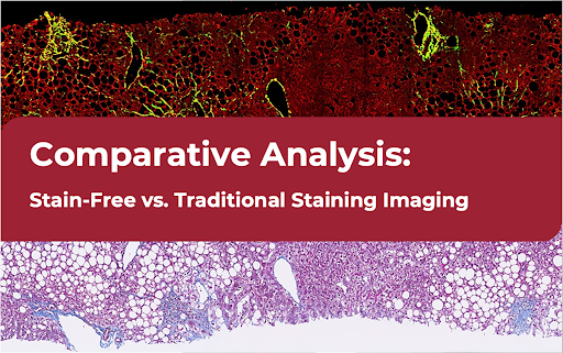
Comparative Analysis: SHG Stain-Free vs. Traditional Staining
Imaging Quality: Accuracy, Consistency, and Reproducibility
One of the key advantages of SHG imaging over traditional staining methods is consistency and reproducibility. Traditional staining may produce inconsistent results depending on variables such as stain quality, application method, and technician expertise. SHG imaging, by contrast, is automated and provides uniform results.
In addition, SHG imaging delivers clearer, more precise images of collagen structures, which are essential in assessing fibrosis. Traditional stains like Masson’s trichrome can stain positively for collagen but may lack the specificity and detail that SHG imaging offers¹².
Time Efficiency and Workflow
Traditional staining involves multiple steps, from tissue preparation to staining and finally digitization, making it a time-consuming process. Each step requires manual effort, and the variability in the staining process can lead to delays¹³. SHG stain-free imaging, on the other hand, streamlines this workflow.
However, it’s important to note that the image acquisition time for SHG-TPE scanning can take 2-3 hours depending on tissue size, The real time-saving comes from the consistency and automation SHG provides—once the images are acquired, they are ready for analysis, reducing the manual labor associated with traditional stains¹⁵.
Accuracy and Reproducibility
Accuracy in diagnostics is paramount, and SHG imaging excels in providing quantitative, reproducible results. Traditional staining is often subjective and can vary between labs. SHG imaging, combined with AI-enhanced analysis, delivers objective data, allowing for more precise diagnostic decisions¹⁸. The ability to accurately visualize collagen structures in fibrosis assessment is one of the main reasons why SHG is increasingly relevant in liver disease research¹⁹.
Additionally, the integration of AI in SHG imaging offers an enhanced level of precision. AI algorithms can detect and quantify fibrosis-related structures automatically, removing potential human error in interpretation²⁰.
However, it’s important to note that the image acquisition time for SHG-TPE scanning can take 2-3 hours depending on tissue size, The real time-saving comes from the consistency and automation SHG provides—once the images are acquired, they are ready for analysis, reducing the manual labor associated with traditional stains¹⁵.
Time and Resource Efficiency
When comparing the overall workflow, SHG imaging reduces the manual effort involved in traditional staining methods. While the acquisition time for SHG images may be similar to the preparation and staining time for traditional methods, SHG’s automation and AI integration help eliminate the time-consuming steps of staining and human-based image interpretation²¹.
With HistoIndex’s Genesis® 200 platform, the imaging process becomes highly efficient, producing consistent, stain-free results with minimal manual intervention, thus speeding up research processes and improving scalability²².
Advantages of SHG Imaging in Liver Disease Research
In liver disease research, detecting fibrosis early and accurately is crucial for developing effective treatments. SHG imaging, with its high precision and ability to capture collagen structures, provides a significant advantage over traditional staining methods. Its application in diseases like MALFD and MASH allows for better disease characterization, making it a valuable tool in both research and clinical settings²⁶.
Several studies have demonstrated the effectiveness of SHG imaging in fibrosis assessment, showing that it offers a more comprehensive understanding of disease progression, which is essential for drug development and treatment planning²⁷.
The Future of Medical Imaging: Moving Toward SHG Stain-Free Solutions
As AI technology continues to advance, the capabilities of SHG imaging will only grow stronger. SHG combined with AI-powered quantitative analysis opens new possibilities for diagnostics and research, allowing for more detailed, data-driven insights²⁸. The future of medical imaging is moving towards innovative, automated solutions, providing faster, more accurate results that will transform how diseases like fibrosis and cancer are diagnosed and treated²⁹.
Stain-Free Imaging With Histoindex
Discover how HistoIndex’s SHG stain-free imaging solutions can enhance your fibrosis research or clinical diagnosis. For more information or to schedule a consultation, please contact us. We build strong foundations of care and trust with our partners. If you have any questions about our stain-free imaging, our team will return your message within 3 business days.
Stain-Free VS Traditional Staining Imaging FAQs
- SHG imaging does not require the use of dyes or chemicals, while traditional staining relies on chemical stains like H&E or Masson's trichrome³.
SHG provides precise imaging of collagen structures, crucial for fibrosis assessment, which is a key factor in liver disease progression¹⁰.
SHG imaging offers greater accuracy and consistency, especially in detecting fibrosis¹¹.
While SHG scanning takes time, the elimination of staining preparation and the automated nature of SHG imaging save significant manual effort¹⁴.
SHG is applicable in other fibrosis-related conditions and in cancer research¹².
The reduction in chemicals, dyes, and manual labor makes SHG more cost-effective²⁴.
AI improves diagnostic precision by automating image analysis and providing quantitative data²⁰.
SHG requires specialized equipment, and its benefits outweigh this initial cost in terms of efficiency and accuracy²³.
While SHG is highly effective, traditional staining still plays a role in certain diagnostic scenarios. However, SHG is rapidly becoming a preferred method in many areas of research²⁹.
Institutions focused on fibrosis, cancer, and liver disease research, as well as pharmaceutical companies conducting drug development, can benefit greatly from SHG technology²⁶
References
- “Histopathology in Medical Research,” Journal of Clinical Pathology.
- “Traditional Staining Techniques: H&E and Masson’s Trichrome,” ScienceDirect.
- “Fibrosis Imaging with Masson’s Trichrome,” Liver International.
- “Limitations of Traditional Staining Methods,” Nature Reviews Pathology.
- “Time and Labor in Histopathological Staining,” Annals of Diagnostic Pathology.
- “Chemical Use and Sample Quality in Traditional Staining,” Laboratory Investigation.
- “Introduction to SHG Imaging Technology,” Optical Microscopy Review.
- “Stain-Free SHG Imaging in Fibrosis Research,” Hepatology.
- “Quantitative Imaging of Tissue Structures with SHG,” PLOS ONE.
- “SHG Applications in NAFLD and MASH Research,” Journal of Hepatology.
- “Comparison of Traditional and SHG Imaging in Diagnostics,” Biomedical Optics Express.
- “Visualizing Collagen Structures with SHG vs. Traditional Staining,” American Journal of Pathology.
- “Workflow Efficiency in Histopathology: Traditional vs. SHG,” Diagnostic Pathology.
- “SHG-TPE Scanning and Image Acquisition Times,” Journal of Microscopy.
- “Automation in SHG Imaging for Diagnostic Consistency,” Digital Pathology Journal.
- “Cost Analysis of Traditional Staining in Medical Labs,” Clinical Laboratory Science.
- “Cost Benefits of SHG Imaging in Large-Scale Studies,” European Journal of Histopathology.
- “SHG Imaging and AI in Diagnostic Precision,” AI in Medicine.
- “SHG’s Role in Fibrosis Assessment,” Fibrosis Research Quarterly.
- “AI-Assisted SHG Imaging for Collagen Quantification,” Nature Biomedical Engineering.
- “Time and Labor Comparisons: SHG and Traditional Staining,” Pathology Research Journal.
- “Efficiency of the Genesis Platform in SHG Imaging,” HistoIndex Publications.
- “Resource Management in Histopathology Labs: Traditional Staining vs. SHG,” Journal of Laboratory Medicine.
- “Scalability of SHG Imaging for Large Clinical Trials,” Liver Research and Diagnosis.
- “SHG Imaging for Extensive Tissue Sample Analysis,” Analytical Cellular Pathology.
- “Advantages of SHG Imaging in Liver Disease Research,” World Journal of Gastroenterology.
- “Case Studies in SHG Imaging for Fibrosis Detection,” Hepatic Research Journal.
- “The Future of AI and SHG Imaging in Medical Diagnostics,” Future Healthcare Journal.
- “The Shift to SHG Stain-Free Solutions in Histopathology,” Trends in Medical Technology.

